
Page 93 - Remedial Andrology
P. 93
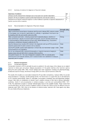
8.2.2.1   Summary of evidence for diagnosis of Peyronie’s disease

             Summary of evidence                                                            LE
             Ultrasound (US) measurement of plaque size is inaccurate and operator dependent.  3
             Doppler US may be required to assess penile haemodynamic and vascular anatomy.  2a
             Intracavernous injection method is superior to other methods to provide an objective assessment of   4
             penile curvature with an erection.

            8.2.2.2   Recommendations for diagnosis of Peyronie’s disease


             Recommendations                                                        Strength rating
             Take a medical and sexual history of patients with Peyronie’s disease (PD), include duration  Strong
             of the disease, pain on erection, penile deformity, difficulty in vaginal/anal intromission due
             to disabling deformity and erectile dysfunction (ED).
             Take a physical examination, including assessment of palpable plaques, stretched or   Strong
             erect penile length, degree of curvature (self-photography, vacuum-assisted erection test
             or pharmacological-induced erection) and any other related diseases (e.g., Dupuytren’s
             contracture, Ledderhose disease) in patients with PD.
             Use the intracavernous injection (IC) method in the diagnostic work-up of PD to provide an   Weak
             objective assessment of penile curvature with an erection.
             Use the PD specific questionnaire especially in clinical trials, but mainstream usage in daily  Weak
             clinical practice is not mandatory.
             Do not use ultrasound (US), computed tomography or magnetic resonance imaging to   Weak
             assess plaque size and deformity in everyday clinical practice.
             Use penile Doppler US in the case of diagnostic evaluation of ED, to evaluate penile   Weak
             haemodynamic and vascular anatomy, and to assess location and calcification of plaques,
             especially prior to surgery.


            8.2.3    Disease management
            8.2.3.1   Conservative treatment
            Conservative treatment of PD is primarily focused on patients in the early stage of the disease as an adjunct
            treatment to relieve pain and prevent disease progression or if the patient declines other treatment options
            during the active phase [995, 1002]. Several options have been suggested, including oral pharmacotherapy,
            intralesional injection therapy, shockwave therapy (SWT) and other topical treatments (Table 26).

            The results of the studies on conservative treatment for PD are often contradictory, making it difficult to provide
            recommendations in everyday, real-life settings [1016]. The Panel does not support the use of oral treatments
            for PD including pentoxifylline, vitamin E, tamoxifen, procarbazine, potassium para-aminobenzoate (potaba),
            omega-3 fatty acids or combination of vitamin E and L-carnitine because of their lack of efficacy (tamoxifen,
            colchicine, vitamin E and procarbazine) or evidence (potaba, L-carnitine and pentoxyfilline)  [1002, 1017-
            1019]. This statement is based on several methodological flaws in the available studies. These include their
            uncontrolled nature, the limited number of patients treated, the short-term follow-up and the different outcome
            measures used [1020, 1021]. Even in the absence of adverse events, treatment with these agents may delay
            the use of other efficacious treatments.


            92                                                SEXUAL AND REPRODUCTIVE HEALTH - MARCH 2021